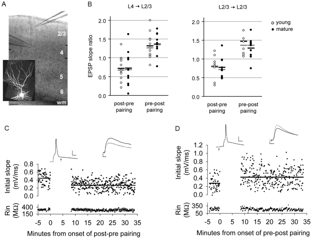
Figure 1. STDP is inducible in mouse primary visual cortex before and after the onset of the critical period of OD plasticity.
(A) Position of field stimulation electrode in layer 4 and whole-cell recording electrode in layer 2/3 in V1, scale bar: 100 microns. WM: white matter. Inset: an alexa-594-filled L2/3 pyramidal neuron. (B) Mean (black bar) and median (gray bar) EPSP slope ratios for young (open circle, LTD n = 12, LTP n = 11) and mature (closed circle, LTD n = 12, LTP n = 11) age groups at vertical layer 4→ layer 2/3 connections (left) and layer 2/3→ 2/3 connections (right). (C) Example of an individual cell in which the EPSP followed the action potential by 9 ms, age = P17. (D) Example of an individual cell in which the EPSP preceded the action potential by 9 ms, age = P17, scale bar 25 mV, 5 ms. Baseline traces are represented by solid lines, post-pairing traces are represented by dashed lines, scale bar: 0.5 mV, 10 ms.

