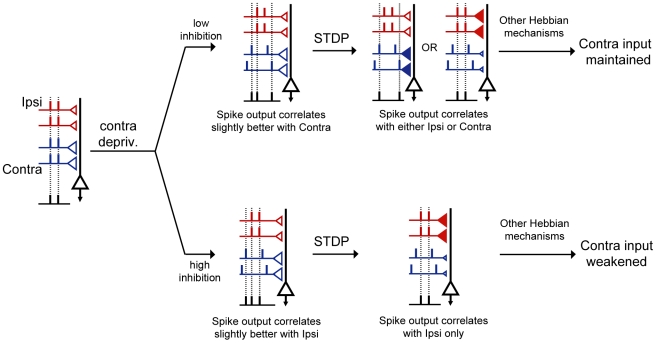
Figure 9. Proposed mechanism by which maturation of inhibition promotes OD shift of class 2/3 neurons in V1.
Symbol designs are the same as in Figure 2. In addition, the filled red and blue triangles highlight the increase of synaptic weights. From left to right, a class 2/3 binocular neuron is driven by convergent ipsi- (red) and contralateral (blue) input pathways. The contralateral bias is indicated by the larger triangles in the blue pathway. Contralateral deprivation decreases the temporal coherence among inputs of the contralateral pathway. The effect of this altered temporal structure on the correlation between pre- and postsynaptic spike times is dependent on the level of inhibition. When inhibition is immature (low inhibition, upper row), spike output of the class 2/3 neuron correlates slightly better with the contralateral input compared to the ipsilateral input. Through STDP, either the ipsi or the contralateral pathway can increase its correlation with the postsynaptic outputs at the expense of a decreased correlation of the other pathway (STDP-mediated increases in synaptic weight are indicated by filled triangles and decreases indicated by smaller open triangles). Since P1 inputs fail to drive postsynaptic spiking, Hebbian mechanisms will not be able to weaken the contralateral deprived eye inputs and strengthen the ipsilateral open eye input. A shift in OD fails to occur. When inhibition is mature (lower row), the spike output of the class 2/3 neuron correlates slightly better with the more coherent ipsilateral, open eye input. In the presence of STDP, open-eye inputs will be able to control postsynaptic spike timing, thus, association-based Hebbian mechanisms can proceed.

