
Fig. 2.
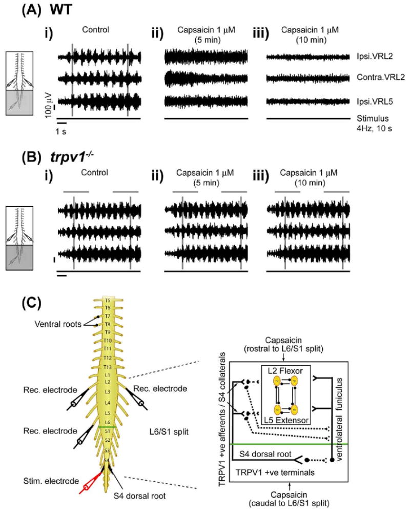
Capsaicin modulation of SCA-evoked locomotor rhythm via TRPV1. Stimulation of the S4 dorsal root can evoke alternating rhythmic discharge from L2 and L5 ventral roots in wild type and trpv1−/− isolated spinal cord preparations, transected at T5. A split-bath was built with Vaseline walls between S1 and L6. Capsaicin was bath applied to the caudal compartment (S1 to cauda equina) of the split bath (grey shaded area). All neurograms were recorded from right L2 (Ipsi. VRL2), left L2 (Contra. VRL2) and right ventral L5 roots (Ipsi. VRL5). Ipsilateral (Ipsi.) and contralateral (Contra.) are abbreviations used for ventral root recordings relative to the side of dorsal root stimulation. The line below each set of neurograms indicates the duration of the train of stimuli applied to the S4 dorsal root (4 Hz, 10 s, 2 T). The voltage and time scales shown in data set Ai are common to all traces in the figure. Vertical grey lines highlight alternating rhythmic discharge of the neurograms. Horizontal grey lines highlight selected number of bursts within the neurogram. (A) Effect of caudal application of capsaicin in wild type preparations. (i) Rhythmic neurogram discharges in untreated control conditions. (ii) Uncoordinated neurogram discharge within 5 min of capsaicin (1 μM) application to the caudal compartment. (iii) Complete blockade of ventral root neurogram discharges following 10 min of continued capsaicin (1 μM) application in the same preparation (n=5). (B) Effect of caudal application of capsaicin in trpv1−/− preparations. (i) Rhythmic neurogram discharges in untreated control conditions. (ii) No change in neurogram discharges within 5 min of capsaicin (1 μM) application to the caudal compartment. (iii) Lack of blockade of neurogram discharges following 10 min of capsaicin (1 μM) application in the same preparation (n=3). (C) Schematic diagram representing the neonatal mouse spinal cord preparations used. There is the stimulation electrode (red) at dorsal S4 root (black) and recording electrodes (black) at right and left L2 and right L5 right ventral roots. A split-bath was built with a Vaseline wall (green) between the L6 and S1 segments. The inset schematic presents the hypothesis that capsaicin application to the caudal compartment of L6/S1 split targets either sacral capsaicin sensitive TRPV1 positive afferent terminals projecting onto lumbar motor networks by way of intercalated interneurons in the VLF or TRPV1 positive S4 afferent collaterals from sacral segments onto lumbar segments via dorsolateral tracts. Capsaicin application to the rostral compartment of the L6/S1 split bath targets TRPV1 positive S4 afferent collaterals originating from sacral segments onto lumbar segments. These TRPV1 positive afferents can rostrocaudally modulate lumbar locomotor CPG networks which may include polysynaptic projections onto the VLF. For interpretation of the references to color in this figure legend, the reader is referred to the Web version of this article.