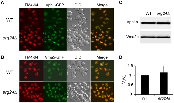
Figure 3. Expression and localization of V-ATPase in erg24Δ.
Vph1p (A) and Vma5p (B) were tagged with C-terminal GFP at their chromosomal loci in WT and erg24Δ strains. Vacuolar membranes were stained with FM4-64 for 30 min in YPD and chased for another 30 min with fresh YPD. (C) Immunoblotting of Vph1p and Vma2p in vacuolar vesicles isolated from WT and erg24Δ. (D) Vma2p to Vph1p ratio in WT was designated as V1/Vo ratio of 1. Calculation was based on data from three independent vacuolar vesicle preparations for each strain. Means of the ratios and standard deviation are plotted.

