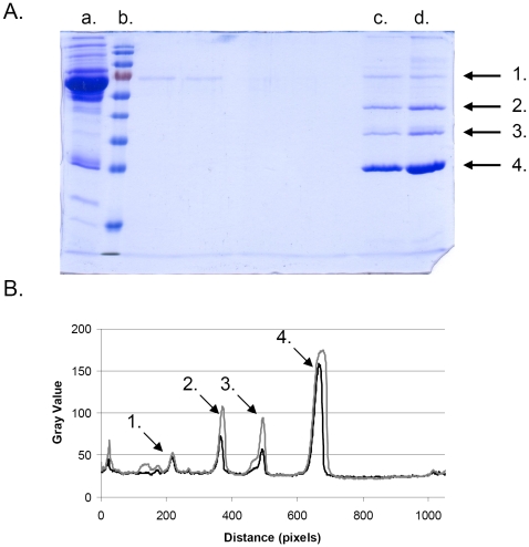
Figure 4. Hard corona around the copolymer particles after 30 s and 6 h of incubation in human plasma.
A) 12% SDS-gel showing; a: human plasma diluted 20 times (5 µl added to the gel), b: MW standard, c: copolymer particles incubated 30 s in plasma, d: copolymer particles incubated 6 h in plasma. Arrow 1–4 indicates the gel bands for serum albumin, apolipoprotein A-IV, apolipoprotein E, and apolipoprotein A-I, respectively. B) traces for lane c and d in panel A as obtained using ImageJ (see the Materials and Methods section). The arrows indicate the same proteins as in panel A.

