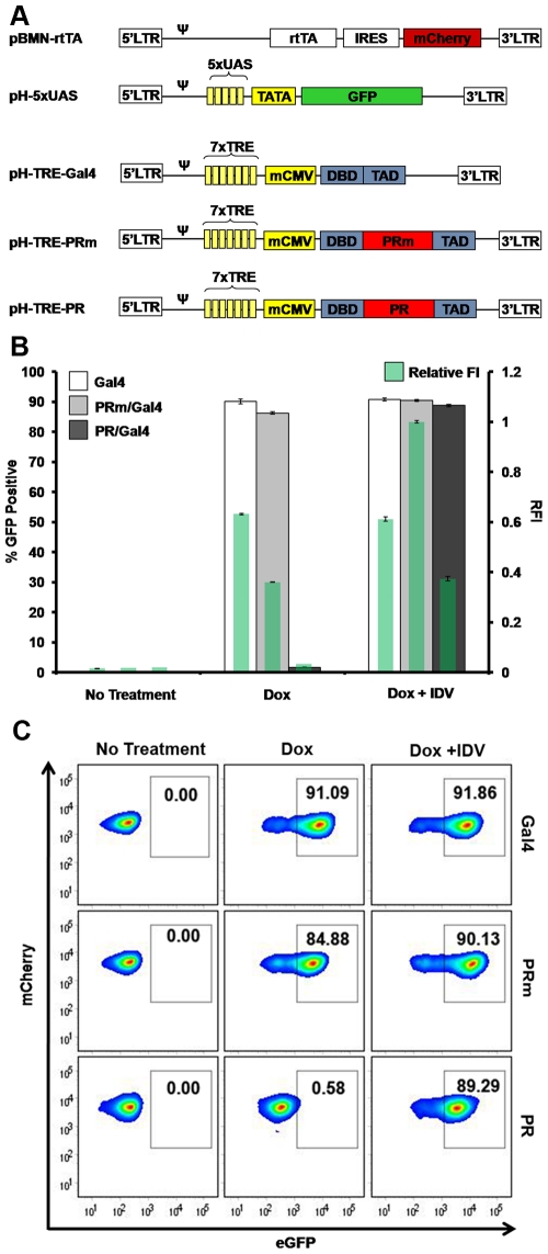
Figure 3. Establishment of a clonal T-cell line stably expressing inducible assay elements.
(A) Constructs utilized to generate infectious particles for the transduction of SupT1 cells with the various assay elements (the rtTA, eGFP reporter, and Gal4-based vectors: Gal4, PR/Gal and PRm/Gal4). (B) Flow cytometry analysis of selected clones. Clones expressing the elements of the assay were analyzed with no treatment, with 1µg/mL Dox, or with 1µg/mL Dox and 10µM IDV. (C) Quantification of eGFP expression (left axis and larger bars) and the relative fluorescence intensity (RFI) of each sample (right axis and green bars within larger bars) of clones treated as indicated. The RFI was calculated by normalizing green mean fluorescence intensity (MFI) to the brightest MFI observed (PRm with Dox + IDV).

