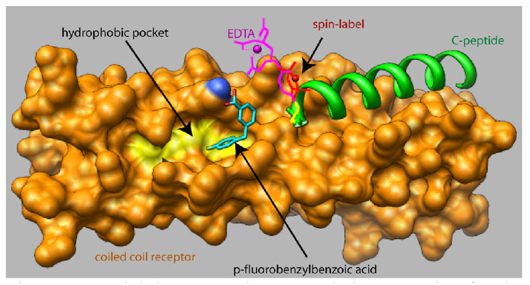
Figure 1.
Model demonstrating second site screening for the hydrophobic pocket. A spin label or EDTA moiety is attached to an outer C-peptide (green) which binds in a site adjacent to the pocket. The coiled coil is shown as an orange surface with the floor of the pocket emphasized in yellow. The small ligand p-fluorobenzylbenzoic acid is shown docked in a pose which agrees with the NMR data (see text).

