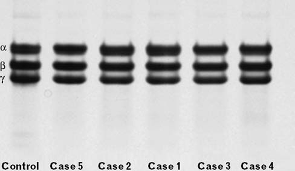
Fig. 1.
Electrophoresis of the fibrinogen on a 6% gel, SDS/PAGE Tricine-system, under reducing conditions. The gel was loaded with 14 µg of non cross-linked fibrin from each sample, prepared as described in the Materials and Methods section. In the control gel lane, the fibrin α, β, and γ chains are indicated on the left-side.

