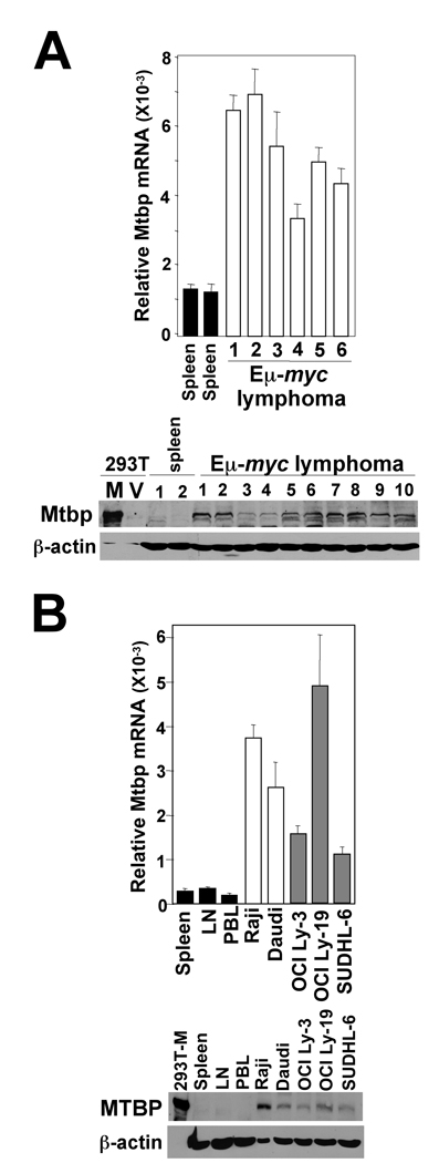
Figure 7.
Increased Mtbp expression in murine and human lymphomas. (A, B) qRT-PCR and Western blots were performed with splenocytes (prior to detectable disease) and B cell lymphomas from wild-type Eµ-myc transgenic mice (A) and human B cell lymphoma lines (Burkitt lymphomas, white bars; DLBCL, grey bars) and normal human lymphatic tissues (spleen; lymph node, LN; peripheral blood lymphocytes, PBL, black bars) (B). Protein lysates from 293T cells transfected with an Mtbp expressing vector (M) or empty vector (V) were controls (1/6 of the amount of protein of the other samples). qRT-PCR data were generated in triplicate and normalized to β-actin levels.

