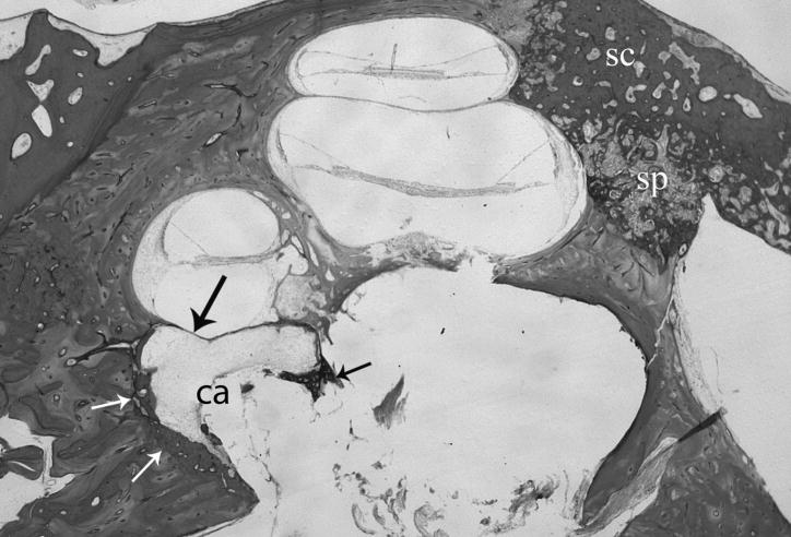
Fig. 1.
Histological section of right temporal bone shows otosclerosis (small arrows) surrounding a cavity (ca) that is continuous with the internal auditory canal and contains some loose fibrous tissue. The process has eroded the endosteal bone leaving only a fibrous membrane between the cochlear basal segment and the cavity (large arrow). (sc) otosclerosis. (sp) otospongiosis. H&E × 15.

