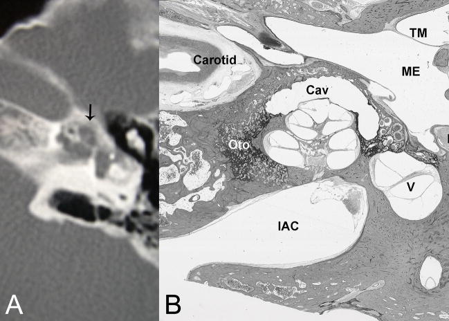
Fig. 3.
(A) CT reveals pericochlear cavities, one of which is in a periapical location. (B) Histological section of right temporal bone of another patient with comparable disease shows substantial otosclerosis foci (Oto) with a large cavity (Cav) capping (anterior and lateral to) the apex of the cochlea. (TM) tympanic membrane, (ME) middle ear, (FN) facial nerve, (V) vestibule, (IAC) internal auditory canal. H&E × 8.

