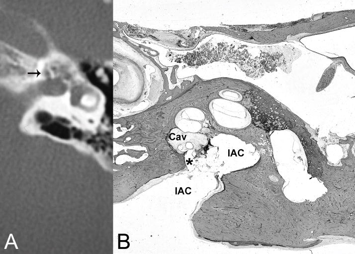
Fig. 4.
(A) CT demonstrates cavitating otosclerosis with pericochlear cavities, one of which communicates with the internal auditory canal near the fundus (arrow). (B) Histological section of right temporal bone of another patient with comparable disease shows otosclerosis cavity (Cav) in continuity (*) with the internal auditory canal (IAC) communicating with the cerebral spinal fluid space. H&E × 8.

