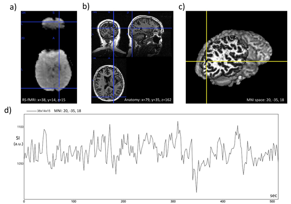
Figure 5.
Voxel time course in right cerebral hemisphere from subject 651. a) Time frame from the resting state fMRI recording, where a voxel-of-interest in the visual area of the occipital lobe is marked with a cross-hair. b) Corresponding voxel in the 3D Tl-weighted anatomical image. c) Same voxel in the volume-rendered 3D anatomical image obtained with MRIcron (http://www.sph.sc.edu/comd/rorden/MRicron) d) Plot of signal intensity (S.I.) versus time for the same voxel.

