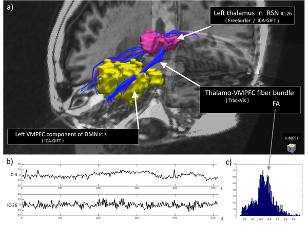
Figure 7.
Integration of structural connectivity (DTI) and functional resting state networks (fMRI). All derived information from the multimodal MRI examination (subj651) was obtained automatically with different freely-available analysis tools. Coregistration was performed in Matlab. a) Tl-weighted anatomical 3-D image superimposed with: (i) anatomical region (thalamus) segmented by FreeSurfer; (ii) resting state networks (RSNs) derived from ICA analysis (GIFT) of the BOLD fMRI recordings; (iii) white matter fiber tracts (TrackVis) between these regions of interest (ROIs). Yellow blob is the ventromedial prefrontal cortex (VMPFC) component of the default mode network (DMN). b) Corresponding time courses (256 frames; TR= 2 s) of the resting state components. Upper trace is IC-3 (DMN). Lower trace is IC-26 located in the central thalamic region (not part of the DMN). c) Histogram of the FA values, reflecting white matter integrity and structural connectivity along the fiber tracts between the two regions: (i) spatial intersection between anatomical thalamus and the RSN defined by IC-26, and (ii) the VMPFC component of DMN defined by IC-3. [Courtesy of Martin Ystad, Tome Eichele, Erlend Hodneland, and Judit Haasz]

