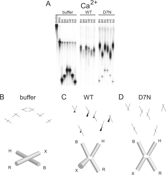
Figure 5.
Conformation of FPV resolvase-HJ complexes analyzed by comparative electrophoresis in the presence of 200 uM CaCl2. A) Comparative gel electrophoresis analysis of fowlpox resolvase-DNA complexes. Markings as in Figure 3. B) Structural interpretation of comparative electrophoresis data for buffer only, C) wild-type FPV resolvase, or D) D7N. Markings as in Figure 3. The continuity of adjacent cylinders seen in the buffer only condition indicates that the adjacent arms of the HJ are coaxially stacked.

