
Figure 4.
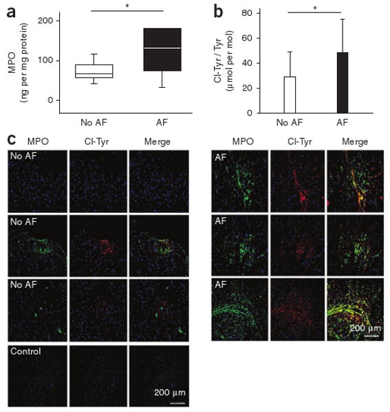
Atrial MPO amounts and protein-bound 3-chlorotyrosine in individuals with or without atrial fibrillation. (a) Quantification of MPO content in tissue homogenates of right atrial appendages of individuals undergoing on-pump coronary bypass surgery, as determined by ELISA normalized to protein concentration (no atrial fibrillation: n = 17, atrial fibrillation: n = 10). Data are presented as median (line) and interquartile range (box); whiskers indicate 5% and 95% percentiles, *P < 0.05. Statistical analyses were performed by the Mann-Whitney U test. (b) LC-MS–based quantitative assessment of 3-chlorotyrosine in atrial tissue of the same individuals. Data are means ± s.d. *P < 0.05. Unpaired Student's t test was used for statistical analysis. (c) Immunofluorescent staining of human right atrial appendage tissue. Left, three representative examples of MPO and the MPO-specific oxidant 3-chlorotyrosine from seven individuals without documented atrial fibrillation. Right, three representative images of right atrial appendage tissue from seven individuals with atrial fibrillation.