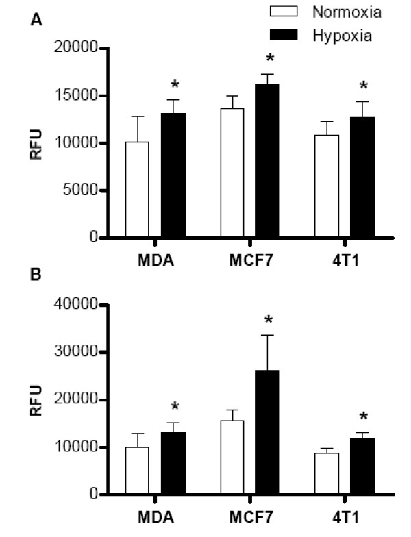
Figure 1.
Migration of breast cancer cells through a microporus membrane (A) and invasion through an extracellular matrix (B) in response to SDF1α in normoxia and hypoxia conditions. Three cell lines MDA-MB-231, MCF7, and 4T1 cells were exposed to either normoxia or hypoxia (2% O2 with 5% CO2) for 48 hrs. Cell migration and invasion were assessed as described in the Methods and expressed as relative fluorescence units (RFU). Data are presented as the mean ± SD of duplicate samples and are representative of three independent experiments. *p < 0.05 versus normoxia-treated cells.

