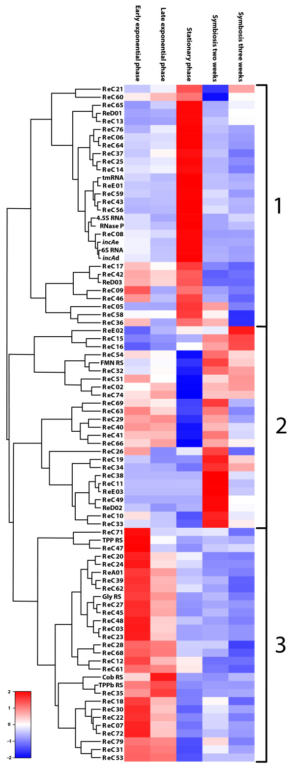
Figure 3.
Heat map of the candidate-ncRNAs. The heat map visualizes the individual ncRNA transcription profiles of the detected ncRNAs that were differentially expressed under our experimental conditions. The expression values in each row were standardized by subtraction of the mean and division by the standard deviation and hierarchically clustered using R. ncRNAs showing similar expression patterns are grouped as follows: group 1, stationary phase; group 2, symbiosis; group 3, exponential phase. The letters b, d and e indicate gene location on the respective plasmids.

