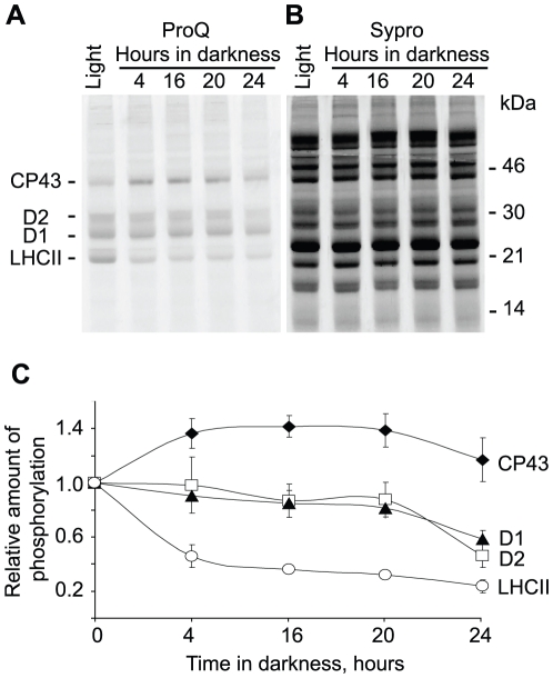
Figure 5. Phosphorylation of thylakoid proteins during prolonged darkness assayed with ProQ phosphoprotein stain.
A, ProQ phospho-stain analysis of SDS-PAGE separated thylakoid proteins from wild type plants exposed to normal light for 4 hours (Light) and then transferred to darkness for 4, 16, 20 and 24 hours, as indicated. B, Sypro total protein stain on the same gel as in A. Positions of the phosphorylated thylakoid proteins and of the molecular mass markers are indicated. C, relative quantification of the protein phosphorylation from the gels stained like in A and B, at the corresponding time points of plant incubation in darkness. Error bars represent S.D. of at least three independent experiments.

