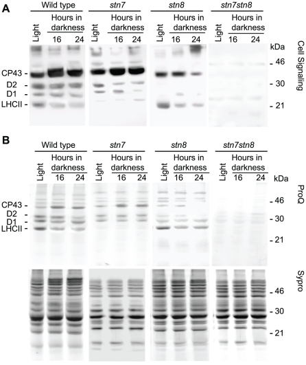
Figure 6. Protein phosphorylation patterns in the stn mutants.
A, immunoblotting analysis of SDS-PAGE separated thylakoid proteins from wild type, stn7, stn8 and stn7stn8 mutant plants, as indicated, with anti-phosphothreonine antibody from Cell Signaling. B, ProQ phospho-stains and Sypro total protein stains, as indicated, of SDS-PAGE separated thylakoid proteins from the same samples as in A. The numbers 16 and 24 correspond to the time of dark incubation in hours.

