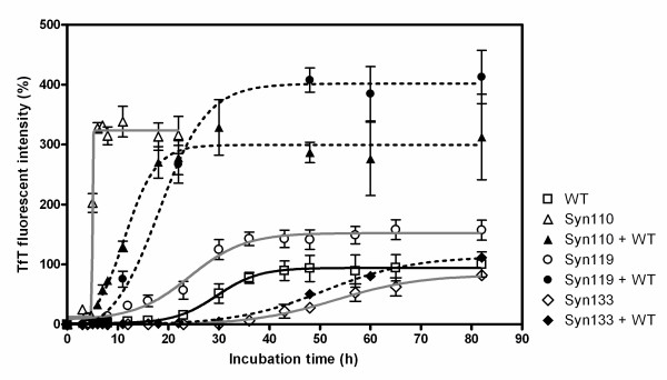
Figure 2.
Fibril formation of C-terminal-truncated variants and the mixture with full-length α-synuclein. The time course of fibril formation was obtained from the TfT fluorescence assay analysis. Seventy μM protein was incubated in triplicate in PBS buffer with 0.02% NaN3, pH 7.4. Full-length α-Syn (white squares), α-Syn110 (white triangles), α-Syn119 (white circles), α-Syn133 (white diamonds), and 35 μM full-length α-Syn + either 35 μM α-Syn110 (black triangles) or 35 μM α-Syn19 (black circles) or 35 μM α-Syn133 (black diamonds).

