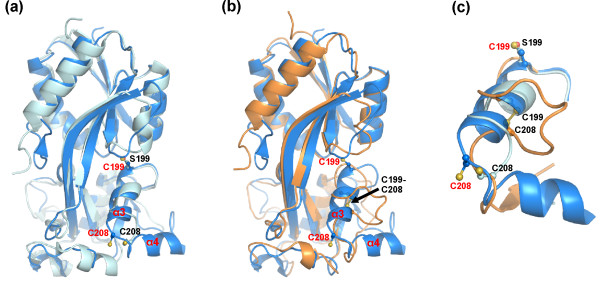
Figure 4.
Structural comparison of the N. meningitidis OxyR and E. coli OxyR. (a) Superimposition of the N. meningitidis OxyR (blue) and the 'reduced' C199S E. coli OxyR (grey, PDB code 1I69). (b) Superimposition of the N. meningitidis OxyR (blue) and the oxidised E. coli OxyR (orange, PDB code 1I6A). (c) Comparison of the redox active centre of, N. meningitidis OxyR (blue); 'reduced' C199S E. coli OxyR (grey); and oxidised E. coli OxyR (orange).

