Figure 6.

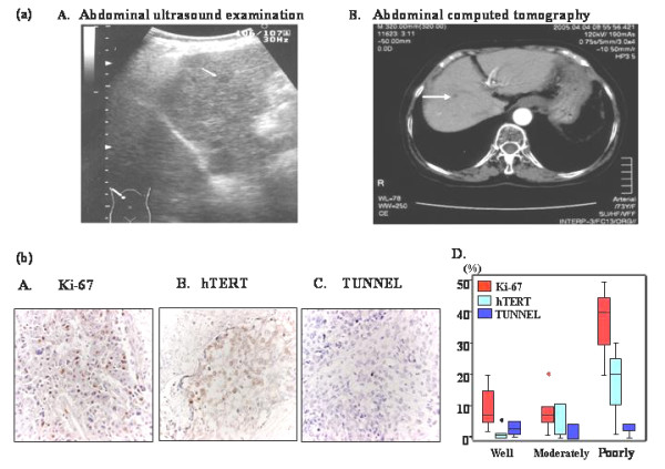
Early detection of small HCC by circulating hTERT mRNA and immunohistochemical analysis of HCC tissues. (a) Imaging diagnosis regarding case with 6 mm HCC. A. Ultrasonography and B. computed tomography of the smallest HCC detected by hTERTmRNA. The diameter of HCC was 6 mm. Left, ultrasonography; right, CT (b) Immunohistochemical analysis of HCC tissues. A. Ki-67 staining (×400), B. hTERT staining (×400), C. TUNEL (×400), D. Labeling indices of Ki-67, hTERT and TUNEL in regard to differential degree of HCC.
