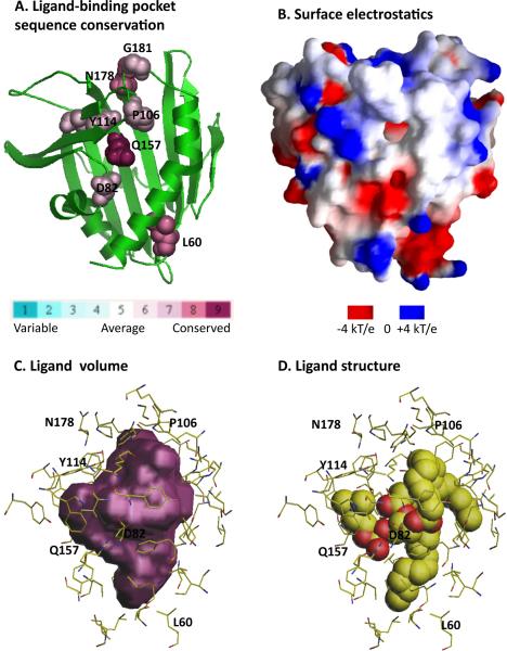
Figure 3.
Analysis of the lipid-binding pocket of the human phosphatidylcholine transfer protein (PC-TP, PDB id 1LN1, [27]). A. Conservation of residues lining the pocket is calculated and displayed by ConSurf [33]. B. Electrostatic surface potential is calculated and imaged with GRASP [34]. C. Volume plot of the ligand binding pocket is generated by VOIDOO [35]. D. The PC-TP ligand DLP (1,2-dilinoleoyl-dn-glycero-3-phosphocholine) is shown in molecular representation. Residues lining the ligand-binding pocket and predicted to be functionally important are delineated in C and D.

