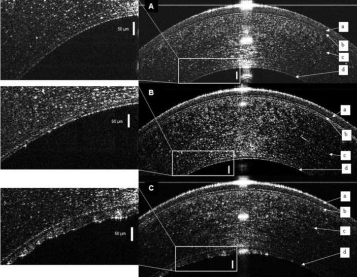
Figure 1. Ultra high resolution optical coherence tomography (UHR-OCT) images of the central cornea.
The epithelium (a), Bowman's layer (b), and stroma (c) are readily seen in each image and Descemet's membrane (d) is enlarged in the inset. (A) Descemet's membrane of a normal young subject appeared as a single smooth opaque line on the back surface of the cornea. (B) Descemet's membrane in a normal elderly subject appeared as a band formed of two smooth opaque lines with a translucent space in between. (C) In UHR-OCT image of the right eye of Fuchs' dystrophy patient number 5, Descemet's membrane consisted of a thickened band composed of two opaque lines separated by a translucent space. The anterior line was smooth while the posterior line had a wavy irregular appearance with areas of localized thickenings. Bars are 50 μm.

