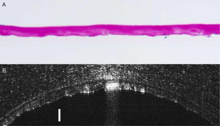
Figure 3. Comparison of corneal histological analysis and ultra high resolution optical coherence tomography (UHR-OCT) images of a Fuchs' dystrophy patient.
Image (A) shows the photomicrograph of the pathology section of the same Descemet's membrane in image (B) obtained by Descemet's stripping automated endothelial keratoplasty (DSAEK) of the right eye of Fuchs' dystrophy patient number 5. Image (A) discloses thickened Descemet's membrane with areas of nodular excrescences correlating with cornea guttae. The average central thickness of the Descemet's membrane measured histopathologically by light microscope was 31μm (Periodic acid-Schiff stain; original magnification X400). UHR- OCT image (B) of the same Descemet's membrane appears as a thickened band formed of two opaque lines. The anterior line is smooth while the posterior line has a wavy irregular appearance with areas of localized thickenings that can be correlated to cornea guttae. In vivo UHR-OCT measurements of this Descemet's membrane was 55 μm. Bar is 50 μm.

