
Figure 5. CRCL induced phosphorylation of NF-κB p65 in vivo.
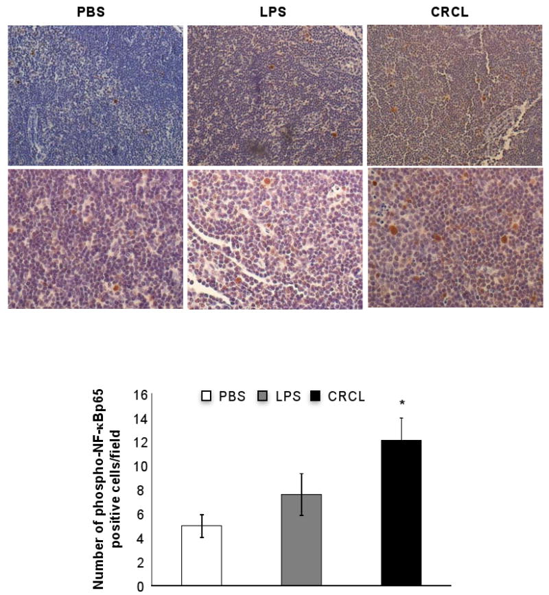
Naïve mice (4 animals per group) were injected (s.c.) with CRCL (100 μl, 25 μg), LPS (100 μl, 1 μg) or PBS as control vehicle. Phospho-NF-κBp65 was detected in the draining lymph nodes by IHC staining. Upper panels, 200× magnification, lower panels 400× magnification. Histogram represents a double blind count of 10 frames of phospho-NF-κB p65 positive cells. *, a significant difference compared to untreated mice (p<0.002).