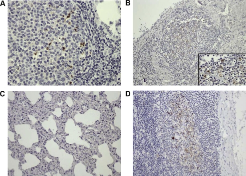
Figure 1.
Detection of PrPSc by immunohistochemistry. (A) Lung, scrapie-VM coinfected animal. Scattered PrPSc deposition within the germinal center of a lymphoid follicle close to an alveolar space, 40×. (B) Mammary gland, scrapie-VM coinfected animal. PrPSc deposition within the germinal center of a lymphoid follicle close to a lactiferous duct, 10×. Insert: Detail of the same image, 60×. (C) Lung, scrapie-VM coinfected animal. Lack of PrPSc detection within the IP, 20×. (D) Mediastinal lymph node, scrapie-VM coinfected animal. PrPSc presence within the germinal centers, 20×. (A color version of this figure is available at www.vetres.org.)

