Figure 4.

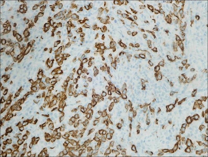
Immunohistochemical stain for pancytokeratin, highlighting tumor cells with unstained lymphocytes in the background (immunoperoxidase stain ×400).

Immunohistochemical stain for pancytokeratin, highlighting tumor cells with unstained lymphocytes in the background (immunoperoxidase stain ×400).