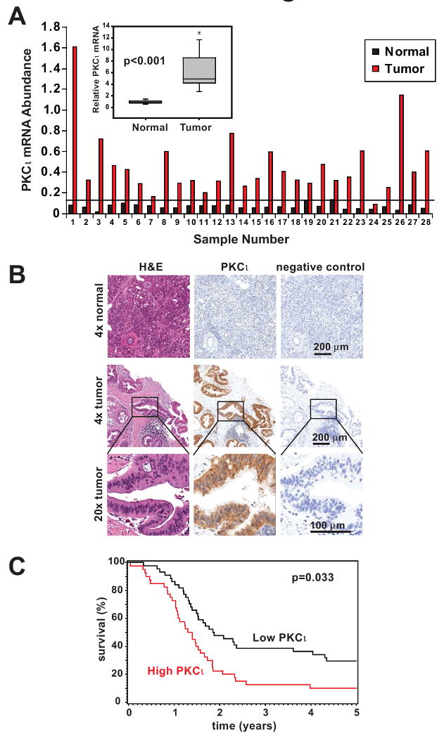
Figure 1. PKCι is highly expressed in human pancreatic cancer and correlates with poor survival in PDAC patients.

A) qPCR analysis of PKCι mRNA expression in 28 matched human pancreatic tumor and adjacent non-tumor pancreas. Data were normalized to 18S RNA abundance (× 104) to control for RNA concentration. Horizontal line indicates 2 standard deviations above the mean PKCι mRNA abundance in adjacent non-tumor pancreas samples. Inset: PKCι mRNA expression is significantly increased in tumors compared to matched non-tumor pancreas tissue. Average fold increase in PKCι mRNA abundance in tumor/matched non-tumor is plotted. B) Representative images of IHC detection of PKCι expression in formalin-fixed human pancreatic adenocarcinoma and normal pancreas. H&E staining and negative control secondary antibody staining are also shown in serial sections. C) Kaplan-Meier survival curves. PDAC patient tumors were analyzed by IHC for PKCι expression and divided into high (red line) and low (black line) expression groups as described in Materials and Methods.
