
Figure 2. PKCι is highly expressed in PDAC cell lines and is not required for anchorage-dependent (non-transformed) growth of PDAC cells.
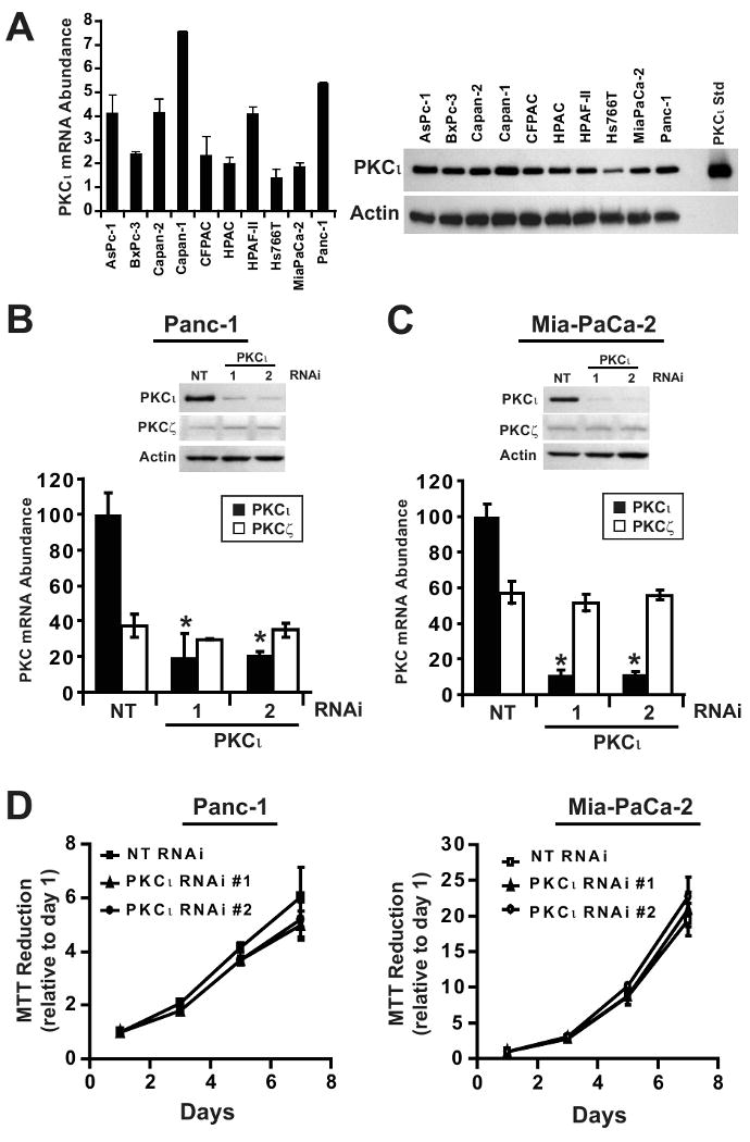
A) Left, qPCR analysis of PKCι mRNA expression in ten human pancreatic cancer cell lines. mRNA abundance is normalized to GAPDH (× 102), n=3. Right, Immunoblot analysis of ten human pancreatic cancer cell lines for expression of PKCι and β-actin. qPCR analysis of PKCι and PKCζ mRNA expression in B) Panc-1 and C) MiaPaCa-2 stably carrying either non target (NT), PKCι-specific RNAi constructs (PKCι #1) or (PKCι #2). PKC mRNA abundance is normalized to GAPDH and presented relative to PKCι in NT RNAi cells. Insets, Immunoblot analysis of PKCι, PKCζ and β-actin protein expression in B) Panc-1 and C) MiaPaCa-2 NT or PKCι-RNAi (PKCι#1 and PKCι#2) constructs. D) Anchorage-dependent growth in Panc-1 (left) and MiaPaCa-2 (right) stably carrying either NT or PKCι-RNAi (PKCι#1 and PKCι#2) was determined by MTT colorimetric assay. Analysis was performed in triplicate and represents two independent experiments.