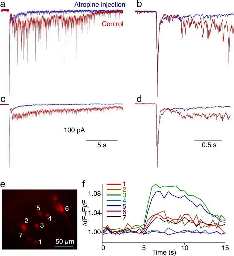
Figure 5. Muscarinoceptive neuron responses to MLR stimulation are inhibited by atropine.
a) Muscarinceptive neurons were targeted for whole cell recordings (labelled retrogradely, see Figs 1–4) and whole cell voltage clamped. Single, high intensity stimuli to the MLR evoked complex EPSCs. Response to a single stimulus is shown in control (red) and atropine (10 μM).
b) Expansion of recording in (a) to demonstrate composition of the late component and to demonstrate the lack of effect of atropine on the early component.
c) Mean response to 5 sequential stimuli in control and atropine.
d) Faster time base of data in (c)
e) Neurons labelled with dye (Alexa 568 dextran, shown and Ca2+ green dextran) by retrograde labelling from dye placed in the MRRN dendritic field.
f) Rise of Ca2+ green dextran fluorescence (Ca2+ concentration) in 7 muscarinoceptive cells when a train of stimulation (5 Hz, 2 s) was applied to the MLR.

