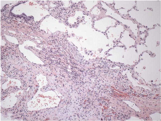
Figure 1. Section of a lung from a patient with idiopathic pulmonary fibrosis.
The section was stained with hematoxylin and eosin. A region of relatively normal lung is at upper right; the main mass of tissue in the center and lower left is fibrotic scar tissue that has replaced normal lung tissue. Figure is courtesy of Dr. Erica Herzog, Yale University School of Medicine, Internal Medicine - Pulmonary and Critical Care Division.

