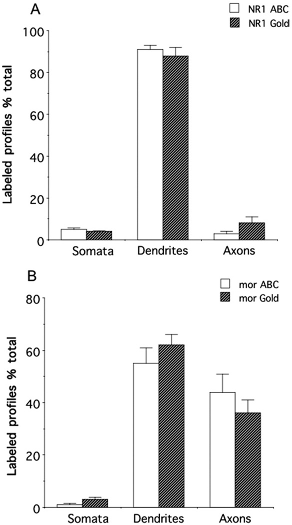
Fig. 6.
Immunolabeling for NR1 or μOR was differentially distributed in neuronal profiles in the CeA. (A) There were similar distribution patterns of NR1 in neuronal profiles when labeled with either avidin-biotin peroxidase complex (ABC) or immunogold (Gold) methods. In particular, there was a high incidence of NR1 in somatodendritic profiles. (B) The neuronal distribution patterns of μOR were in correspondence when labeled with either ABC or immunogold secondary antisera. Immunoreactivity for μOR was present prominently in dendritic and axonal processes.

