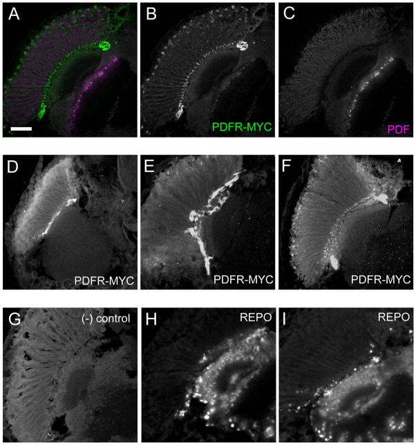
Figure 7. PDFR-MYC in a subset of cells near the fenestrated membrane of the retina.
Adult fly heads sections immunostained showing PDFR-MYC expression near the fenestrated membrane of the retina. (A–C) PDFR-MYC fly heads showing PDF and PDFR-MYC expression. (A) Merged image of (B) (MYC, green) and (C) (PDF, magenta). (D–F) Expression of PDFR-MYC. (G) Negative control: a PDFR-MYC brain stained without incubation in the primary (anti-MYC) antibody. attP2 fly heads with anti-MYC antibody display similar results (not shown). (H–I) anti-REPO staining in w1118 fly heads. REPO is a useful marker for many glial cells: The outer-most layer of REPO-positive cells lies along the fenestrated membrane that separates the retina from the brain: this position is similar to that exhibited by PDFR-MYC positive cells. Scale bar = 50 μm.

