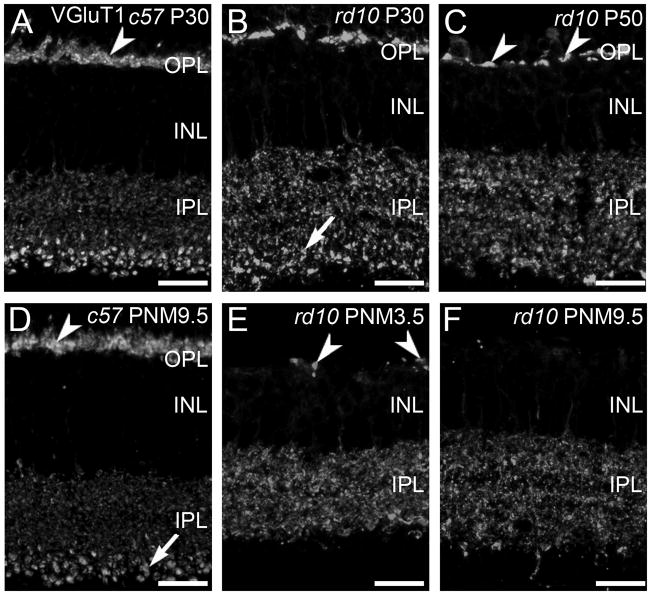
Figure 1. VGluT1 expression in C57BL/6 and rd10 retina.
A, D: In the retinas from wild-type C57BL/6 (c57) mice at postnatal day 30 (P30, panel A) and postnatal month 9.5 (PNM9.5, panel D), VGluT1 antibodies label rod and cone terminals in the outer plexiform layer (OPL) (arrowhead) and bipolar cell terminals in the inner plexiform layer (IPL). Large, lobular rod bipolar cell terminals in the innermost IPL are prominently labeled (arrow). B, C, E, F: VGluT1 expression in the rd10 retina from P30 to PNM9.5. B. At P30, prominent gaps between VGluT1-positive photoreceptor terminals are present in OPL. Rod bipolar cell terminals in the IPL (arrow) are less distinctive than in the wild-type retina. C. At P50, VGluT1 staining in the OPL is reduced (arrowheads). The spacing and morphology of labeled terminals indicates that primarily cone pedicles remain at this time point. E. At PNM3.5, VGluT1 staining in the rd10 OPL labels the few surviving cone terminals (arrowheads). In the IPL, no morphologically distinct rod bipolar cell terminals are discernable. F. At PNM9.5, little to no expression of VGluT1 is detected in the OPL; VGluT1 is still present in the IPL. Scale bar = 20 μm, all panels.

