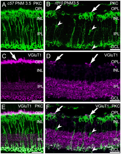
Figure 2. PKCα and VGluT1 double labeling of C57BL/6 and rd10 retinas.
A, C, E: C57BL/6 (c57) retina at PNM3.5. A. Rod bipolar cells labeled with antibodies against protein kinase C-α (PKC) show normal morphology. C. VGluT1 labels photoreceptor terminals in the OPL (arrow) and bipolar cell terminals in the IPL. E. Merged image of PKC and VGluT1 labeling. PKC-positive rod bipolar cell dendrites extend into the plexus of VGluT1-positive photoreceptor terminals in the OPL. Rod bipolar cell terminals in the IPL show strong labeling for VGluT1. B, D, F: rd10 retina at PNM3.5. B. In the rd10 retina, rod bipolar cells have retracted their dendrites and have disorganized axon terminals, with some ectopic processes (arrowheads). Surviving rod bipolar cell bodies are found in clusters (arrows). D. Scattered VGluT1-positive cone terminals remain in the OPL at PNM3.5. F. Merged image of PKC and VGluT1 labeling. Rod bipolar cell bodies cluster near the remaining VGluT1-positive cone terminals in the OPL (arrows). Rod bipolar cell terminals in the inner IPL show reduced labeling for VGluT1, with ectopic processes lacking VGluT1 staining (arrowheads). Scale bar = 20 μm, all panels.

