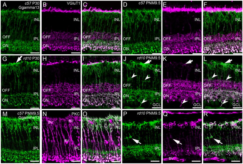
Figure 3. Immunostaining of ON-cone and rod bipolar cells in C57BL/6 and rd10 retinas.
A–C: C57BL/6 (c57) retina at P30. A. Gγ13 labeling of dendrites, cell bodies and axon terminals of all ON-bipolar cells (rod and cone). Large, lobular rod bipolar cell terminals in the innermost IPL are intensely labeled. B. Antibodies against vesicular glutamate transporter 1 (VGluT1) label the axon terminals of rod and cone photoreceptors in the OPL and both ON and OFF bipolar cells in the IPL. C. Overlay of panels A and B showing ON-OFF stratification in the IPL. Terminals of OFF-cone bipolar cells in the distal IPL are VGluT1-positive, Gγ13-negative. Terminals of ON-cone and rod bipolar cells are VGluT1-positive and Gγ13-positive and can be distinguished by size and position in the inner IPL. D–F: C57BL/6 retina at PNM9.5. The labeling patterns for Gγ13 (D) and VGluT1 (E) remains unchanged. F. Overlay of D and E. G–I: rd10 retina at P30. G. Labeling for Gγ13 shows abnormally thickened dendritic processes in the OPL (arrow). ON bipolar cell terminals stratify in the correct sublamina of the innermost IPL. H. A discontinuous band of VGluT1 immunoreactivity is still present in OPL. VGluT1-positive bipolar cell terminals are present throughout the IPL but the rod bipolar terminals are less distinctive than in the wild-type retina. I. Overlay of Gγ13 and VGluT1 labeling shows proper lamination of ON and OFF bipolar cell terminals in the IPL. J–L: rd10 retina at PNM9.5. J. Gγ13-positive dendritic processes are generally absent, although some dense areas of staining persist (arrow). Most ON bipolar cell terminals stratify properly within the ON sublamina of the IPL, although some ectopic processes are present at the INL/IPL border and extend into the ganglion cell layer (GCL) (arrowheads). K. Few sites of VGluT1 labeling remain in the OPL (arrow). Bipolar cell terminals throughout the IPL show VGluT1 labeling but rod bipolar cell terminals are not morphologically identifiable. L. Overlay of Gγ13 and VGluT1 labeling. In the OPL, areas of dense Gγ13 staining are associated with remaining VGluT1-positive puncta (arrow). In the IPL, Gγ13 and VGluT1 co-localize in ON bipolar cell terminals and the ON and OFF stratification remains largely intact. Many ectopic Gγ13-positive processes (arrowheads) also show VGluT1 labeling. M–O: Retinas from C57BL/6 at PNM9.5 immunostained for (M) Gγ13, showing all ON bipolar cells and (N) PKC, showing rod bipolar cells. O. An overlay of M and N distinguishes rod and ON cone bipolar cells. P–R: Retinas from rd10 mice at PNM9.5 immunostained for (P) Gγ13, and (Q) PKC. Ectopic processes in the INL show strong Gγ13 labeling and faint PKC labeling (arrows). R. Overlay of P and Q showing clusters of rod and ON-cone bipolar cells in the INL. The appropriate stratification of rod bipolar and ON-cone bipolar cell terminals in the IPL is still apparent. Scale bar = 20 μm, all panels.

