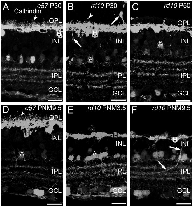
Figure 4. Calbindin immunostaining in C57BL/6 and rd10 retinas.
A, D: C57BL/6 (c57) retina at P30 (A) and PNM9.5 (D). Antibodies against calbindin label horizontal cells and their fine processes associated with the terminals of photoreceptors (arrowheads) in the OPL and a subset of amacrine cells and their terminals in the INL and IPL. B, C, E, F: rd10 mouse retina. B. At P30, during early stages of degeneration, puncta associated with horizontal cell dendrites and axon terminals are reduced but still present (arrowhead). Some horizontal cells sprout ectopic processes (arrows). C. At P50, the puncta associated with fine processes of horizontal cells are no longer detected. E. At PNM3.5, staining of horizontal cell lateral processes in OPL is greatly reduced. F. At PNM9.5, gaps are visible in the calbindin-positive horizontal cell plexus in the OPL. Ectopic processes from horizontal cells are still detected (arrows). Amacrine cells show minimal changes and the stratified organization of amacrine cell processes in the IPL is preserved. Scale bar = 20 μm, all panels.

