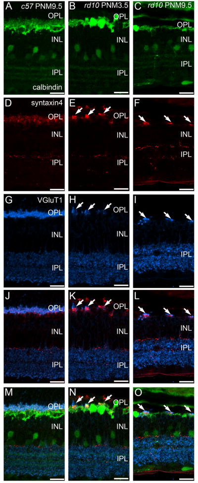
Figure 5. Immunostaining for calbindin, syntaxin 4 and VGluT1 in the C57BL/6 and rd10 retinas at late stages of degeneration.
A, D, G, J, M: C57BL/6 (c57) retina at PNM9.5. A. Calbindin labeling. D. Syntaxin 4 labeling of the fine dendrites of horizontal cells and a sparse plexus in the IPL. G. VGluT1 labeling. J. Overlay of panels D and G showing the direct apposition of presynaptic VGluT1 expression with postsynaptic syntaxin 4 expression in the OPL. M. Overlay of panels A, D, and G showing the normal relationship of presynaptic VGluT1 and syntaxin 4 in fine horizontal cell processes and the remainder of the horizontal cell plexus in the OPL. B, E, H, K, N: rd10 retina at PNM3.5. B. Calbindin labeling reveals horizontal cell process loss. E. Syntaxin 4 labeling in the OPL is restricted to small, widely spaced patches (arrows), but remains essentially normal in the IPL. H. Restricted sites of VGluT1 labeling in the OPL (arrows). K. Overlay of panels E and H showing direct apposition of syntaxin 4 and VGluT1 labeling in the OPL. N. Overlay of panels B, E, and H, shows the relationship of presynaptic VGluT1, syntaxin 4 and the horizontal cell plexus in the OPL. C, F, I, L, O: rd10 retina at PNM9.5. C. Calbindin labeling reveals a severe reduction of horizontal cell processes at PNM9.5. F. Patches of syntaxin 4 labeling in the OPL become smaller at PNM9.5 in the rd10 retina (arrows). I. Surviving cone terminals in the PNM9.5 rd10 OPL express VGluT1. L. Overlay of panels F and I showing juxtaposition of syntaxin 4 and VGluT1 labeling in the OPL. O. Overlay of panels C, F, and I, showing the close physical relationship between syntaxin 4 expression, the horizontal cell plexus and surviving terminals of cones in the OPL at late stages of degeneration. Scale bar = 20 μm, all panels.

