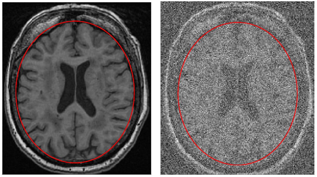
Fig. 1.
The image on the right is the result of applying heavy white noise to the same image shown on the left. The ellipse on the left image is a region-delineation that partitions the image into brain region(inside the ellipse) and the non-brain region(outside of the ellipse). The ellipse on the right is the same as that on the left but due to the noise, it is no longer clear that the ellipse on the right truly does partitions the image correctly. Given the noisy image, different ellipses with different areas seem to fit the brain equally well.

