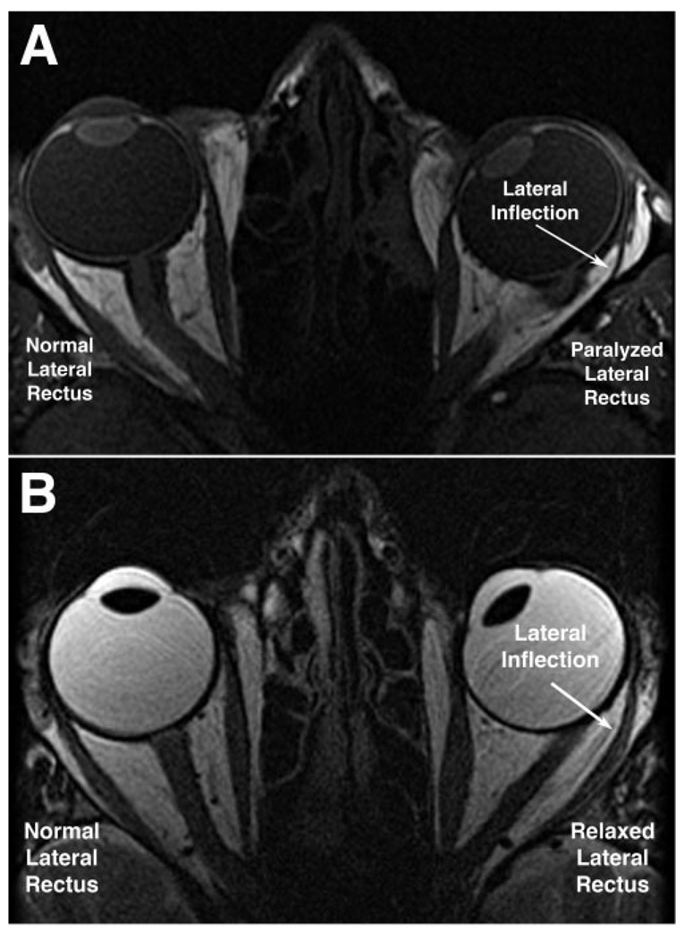
FIGURE 2.
Axial MRI showing LR paths during right eye fixation of a central target. (A) T1-weighted image in subject P1 shows lateral inflection and atrophy of paralyzed left LR muscle during central fixation by the normal right eye. Note left ET, and note that the thinned posterior belly of the left LR is immediately adjacent to the orbital wall. (B) T2-weighted image in subject ET1 exhibiting left ET, similar in magnitude to that of subject P1 with paralytic ET. Note that the relaxed, nonparalyzed LR is thicker and less markedly inflected than the paralyzed LR.

