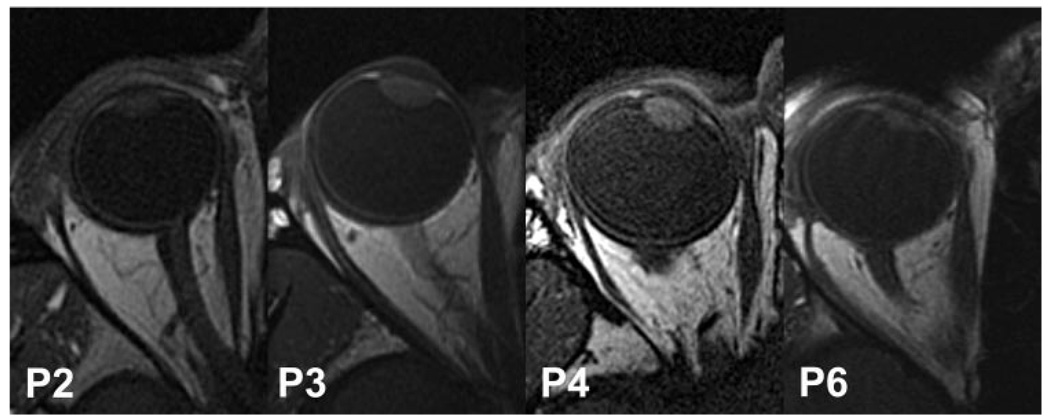
FIGURE 4.
T1-weighted axial MRI of the affected eyes of four subjects with LR paralysis, illustrating the spectrum of lateral inflection of the LR path. Inflection was sharper in subjects P6 and P2 than in subjects P3 and P4. For clarity, all images have been depicted as right eyes, using digital reflection as necessary.

