
Figure 1.
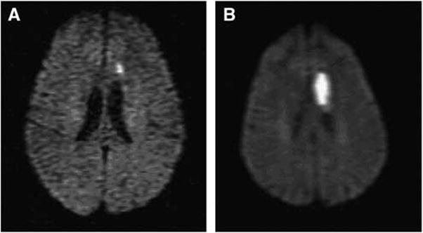
Diffusion weighted imaging of patient with TIA (A) who returned to ED 4 days later with stroke. (B) A 61-year-old woman with history of hypertension and prior basal ganglia lacunar stroke, presented initially with mild expressive aphasia and unsteadiness. Symptoms improved, she was discharged on antiplatelet agent, adjusted antihypertensive regimen, but returned 4 days later with worsened aphasia, right hemiparesis, and slow unsteady gait. Diffusion magnetic resonance imaging initially showed small left frontal subcortical infarction, and repeat scan shows significant extension of stroke in this region.