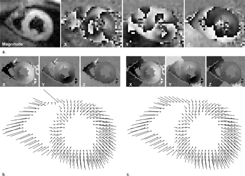
Figure 6.
(a) Magnitude image and initial X, Y, and Z displacement-encoded phase maps. (b, c) Illustrations of phase unwrapping of an end-systolic cine frame of a short-axis view obtained in a volunteer. Spatial resolution is 3.0 mm. In-plane projections of three-dimensional displacement vectors are shown. The X-, Y-, and Z-encoded phase maps are shown at the top of each image. In b, the conventional phase-unwrapping technique leaves a phase fringe (arrows) in the anterior right ventricle. In c, the APU technique removes errors and pushes phase fringes beyond ventricular walls.

