
Figure 6.
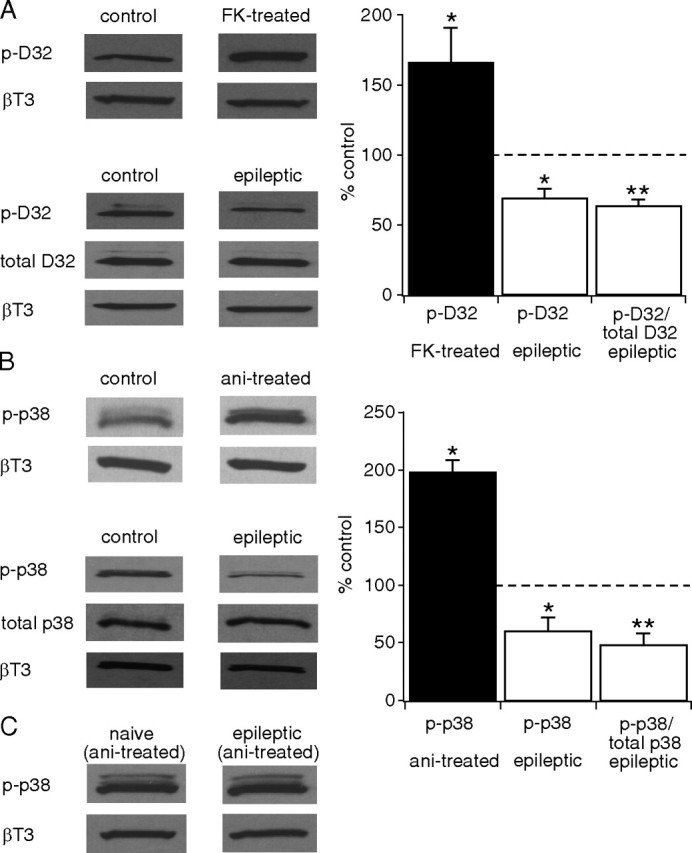
CaN activity is upregulated and p38 MAPK activity is downregulated in the CA1 hippocampal area from epileptic animals. A, Application of FK506 (FK) to hippocampal slices from naive animals increased phosphorylation levels of DARPP-32 (p-D32) compared with untreated slices (control) (*p < 0.05), showing p-DARPP-32 levels are sensitive to dephosphorylation by CaN. p-DARPP-32 levels in CA1 hippocampal tissue were decreased in epileptic animals compared with control, implying increased CaN activity (*p < 0.05). The ratio of p-DARPP-32 to total DARPP-32 was further decreased (**p < 0.01). All p-DARPP-32 and total DARPP-32 levels were normalized to β-tubulin III (βT3) protein levels. Representative Western blots of p-DARPP-32, total DARPP-32 and βT3 protein levels are shown in each condition. Statistical significance compared with control levels. B, Anisomycin (ani) treatment of hippocampal slices from naive animals caused an increase in phosphorylated p38 MAPK (p-p38) levels in the CA1 hippocampal area compared with control (*p < 0.05). p-p38 MAPK levels in CA1 hippocampal tissue were decreased in epileptic animals compared with control (*p < 0.05), implying decreased p38 MAPK activity. The ratio of p-p38 MAPK to total p38 MAPK was further decreased (**p < 0.01). All p-p38 MAPK and total p38 MAPK levels were normalized to βT3 protein levels. Representative Western blots of p-p38 MAPK, total p38 MAPK and βT3 protein levels are shown in each condition. Statistical significance compared with control levels. C, Representative Western blots show that p-p38 MAPK levels following anisomycin treatment of hippocampal slices from both naive and epileptic animals are similar to each other in the CA1 hippocampal area, implying that activation of p38 MAPK cascade by anisomycin is intact in epileptic animals.