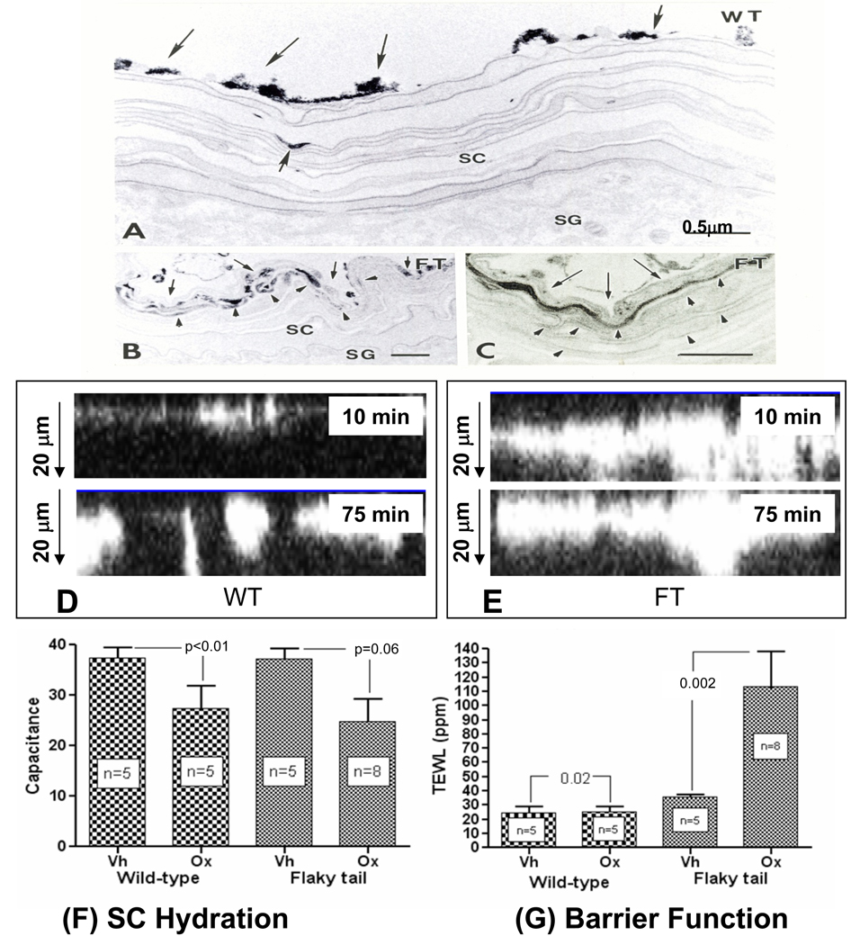
Figure 3. Epicutaneous Tracer Penetrates Flaky Tail Stratum Corneum via the Extracellular Spaces.
A–C: Flanks of flaky tail (FT) and wild-type (WT) mice (n = 3 each) were immersed in 4% lanthanum nitrate solution for 30 min to 2 hrs, followed by aldehyde fixation. After 2 hrs, little or no tracer entered the stratum corneum (SC) of WT mice (A, single small arrow). In contrast, abundant tracer reaches ≥4 layers into the SC in FT mice (B, arrowheads), which on higher magnification [C] can be seen to localize to extracellular domains (C, arrowheads). D+E: Calcium green was applied to freshly-obtained explants from ft/ft and wt mice (n = 3 each), and penetration was assessed by dual-photon confocal microscopy, along the ‘z’ axis after 10 and 75 min. Note much deeper ‘outside-to-inside’ penetration of calcium green in ft/ft mice at both time points. Flaky tail (FT) mice, repeatedly-challenged with low-dose Ox, display a more severe barrier abnormality (G), but no further decline in stratum corneum (SC) hydration in comparison to wild-type (WT) mice (F). A–C: Osmium tetroxide post-fixation; mag bar = 0.5 µm.

