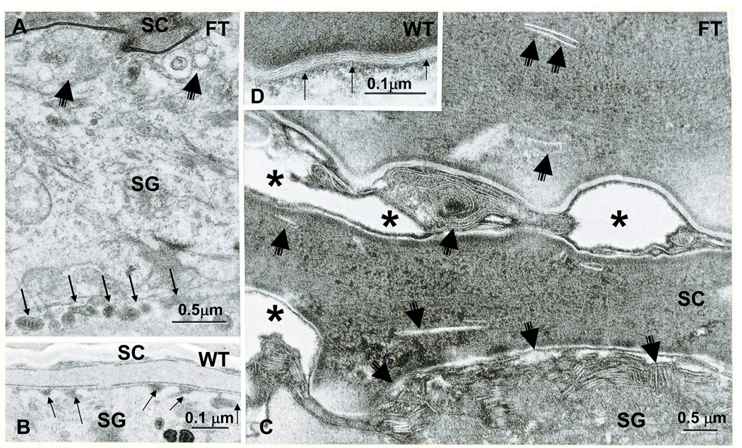
Figure 4. Abnormalities in the Lamellar Body Secretory System in Young Flaky Tail Mice.
Partial failure of lamellar body exocytosis is evident in flaky tail (FT) epidermis (A, C). Note lamellar bodies lined up in peripheral cytosol (A, multiple thin arrows); decreased secreted material at stratum granulosum (SG)-stratum corneum (SC) interface (A&C, short, fat arrows); decreased numbers of lamellar bilayers (C): delayed maturation of lamellar bilayers (C); and entombed lamellar body contents within the corneocyte cytosol (C, short, thin arrows). B&D: Normal lamellar body secretion (B, arrows) and extracellular lamellar bilayers in wild-type (WT) epidermis (D, thin arrows) A&B, osmium tetroxide post-fixation; C&D, ruthenium tetroxide post-fixation. Mag bars = A&C, 0.5 µm; B&D, 0.1 µm.

