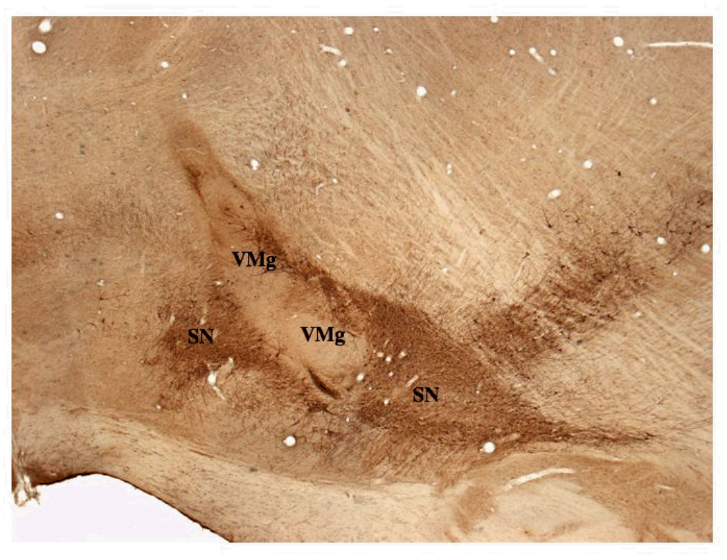
Figure 3.
A. This low power parasagittal view of the rostral brain stem shows the relative position of the VM graft in the SN (VMg) and the host substantia nigra (SN) in a group 1 animal (X200). Rostral is to the left and the injection site for FG is to the left out of the field of view. The VM graft is elongated in the dorso-ventral axis and contains many TH positive neurons which are shown in Figures 4 – 6 at higher power.

