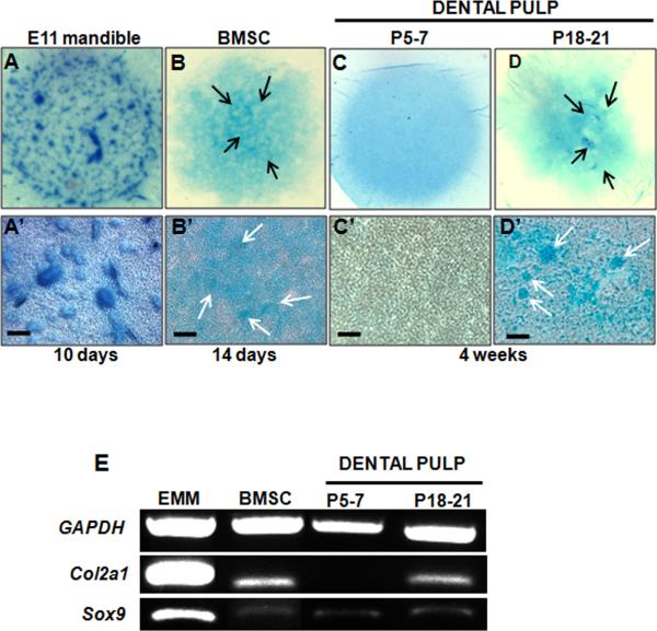
Figure 7. Chondrogenic differentiation by cultures derived from dental pulp.
A-D are representative images from the whole micromass cultures stained with Alcian Blue. Micromass cultures were established from mesenchyme obtained from E11.5 mandibles (A), BMSC (B) and dental pulp obtained from unerupted (P5-7) (C) and erupted (P18-P21) (D) molars. Cultures were grown under chondrogenic condition for 10 days (A), 14 days (B) and 4 weeks (C, D). A’-D’ represent bright field images of the same cultures. Micromass cultures derived from E11.5 mandibular mesenchyme display extensive chondrogenesis indicated by cartilage nodules stained with Alcian Blue (A, A’). Limited number of Alcian Blue stained nodules is detected in micromass cultures derived from BMSC (arrows in B, B’) and dental pulp from erupted (P18-P21) molars (arrows in D, D’). Chondrogenesis and Alcian Blue stained nodules were not detected in micromass cultures derived from dental pulps from unerupted (P5-7) molars (C, C’). Scale bars in A’-D’= 100 μm.
(E) RT-PCR analysis of expression of marker of chondrogenesis in micromass cultures Note the high levels of Col2a1 and Sox9 expression in micromass cultures established from E11.5 mandibular mesenchyme. Levels of Col2a1 expression in the micromass cultures from the erupted (P18-P21) molars and BMSC are low but detectable. Also note the lack of Col2a1 expression in the micromass cultures from the dental pulps from unerupted (P5-7) molars. Sox9 expression was detected in low but detectable levels in all micromass cultures.

